HIV was first identified back in 1980s but scientists are still far from fully understanding it. To get a better notion of how the virus works, a group of scientists have created a detailed image of HIV virus infecting gut tissues.
The image has been created courtesy of biologists from Massachusetts and California. To create it, first the lab mice were transplanted with human bone marrow as well as live and thymus cells. This was done to add such immune cells to the mice’s system which are typically a part of human immune system, and usually attacked by HIV.
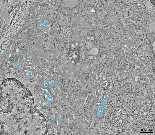
In the next step, these mice were then infected with HIV. The virus was allowed to take its course for 10 to 20 weeks, after which the mice were dissected to examine the damage wrecked by the virus. Using an electron microscope, researchers were able to spot the virus in action in the mice’s gut tissues.
Interestingly enough, rather than attacking the immune cells first, the HIV virus moved away from it and attacked deep inside the intestine. Moreover, another intriguing phenomenon called ‘virological synapse’ was observed in the images taken from the lab mice.
Virological synapse refers to a virus’ ability to spread merely by touching another cell. HIV imitate this behavior and the aforementioned experiment shows that it was able to spread through the gut tissues by touching the neighboring cells. In the image posted above, blue and purple globules can be seen pushing through the gut tissues. These are HIV viruses, embedded deep in living tissues and infecting them.
Source: California Institute of Technology
Courtesy: Pop Sci
[ttjad keyword=”solar-device”]