The first MRI video of childbirth in Germany amazed all. After the success of that childbirth video using Magnetic Resonance Imaging (MRI), a team at Robert Steiner MR Unit of Imperial College London, is using the cinematic MRI to study twin-to-twin transfusion syndrome.
Twin-to-twin transfusion syndrome is something that can generally be fixed with an operation that blocks the offending shared blood vessels. Doctors are sure that it affects the brain development, but they still don’t know exactly how that happens. In twin-to-twin transfusion syndrome, one baby gets excessive supply of blood and the other doesn’t get sufficient blood. Therefore, the growth of the second (who doesn’t get sufficient blood) baby is stunted. This is a disorder between the two babies’ which occurs due imbalance in blood supply.
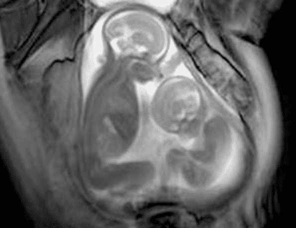
Marisa Taylor-Clarke, a team member of Robert Steiner MR Unit, is using the MRI technique to study a twin-to-twin transfusion syndrome. The usual imaging method takes pictures of little slices of the body, but the cinematic MRIs capture the same slice over and over and pulls them together to make a pretty amazing video. In fact, these snapshots shows us an unprecedented view of inside the womb. Have a look at the video.
In the upper video, we can see the twins’ tiny fists, little feet kicking, and even their brains. If we look carefully, we’ll see the heads are not fully grown yet.
The team of Robert Steiner MR Unit have scanned 24 sets of twins. They found these new pictures (you can say a video of pictures) are letting them see things they’ve never seen before. These pictures offer them important clues like – how to better repair any issues, twin development in general etc. The goal of this technology is to identify developmental problems of the child prior to birth, which can assist parents and doctors with treatment and management strategies.
Source : New Scientists
[ttjad keyword=”camcorders”]


